X-Light V2
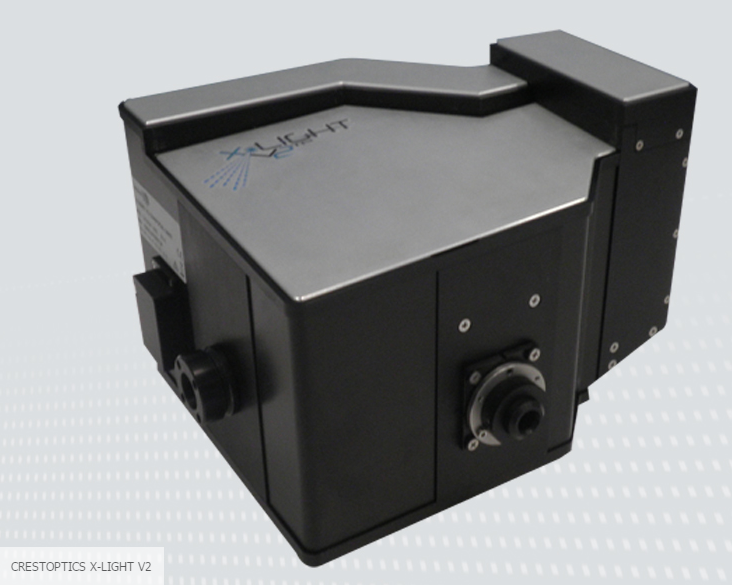
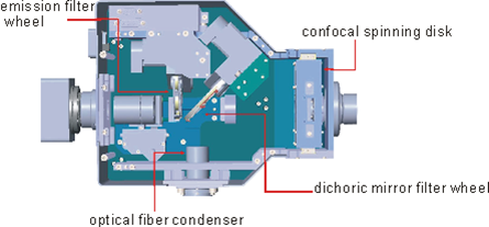
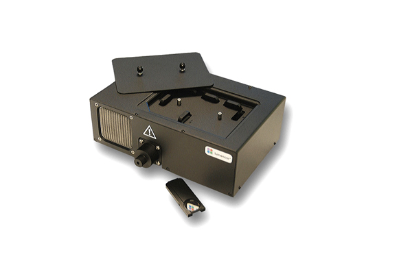
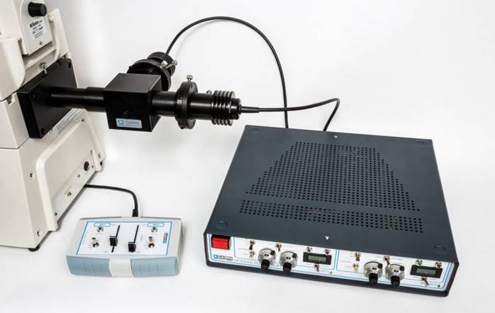
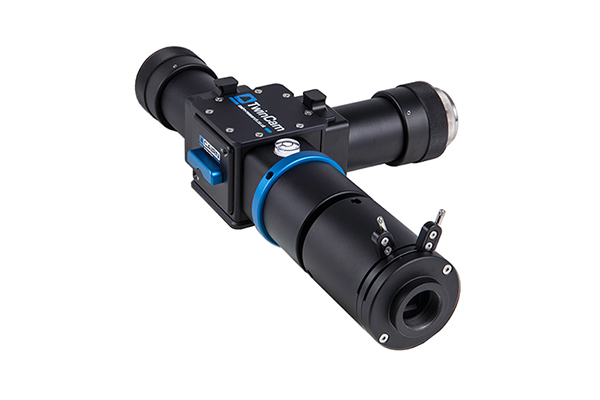
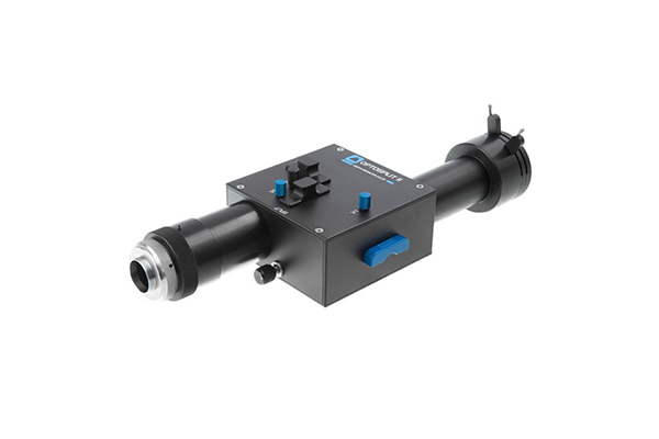
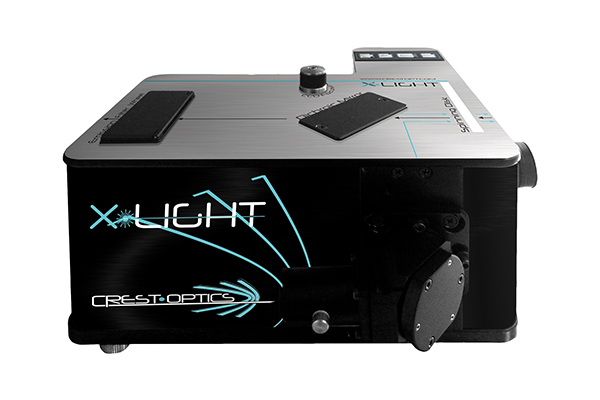
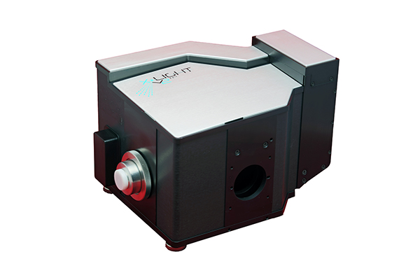
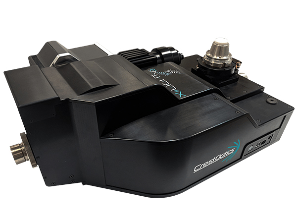
Crest Optics X-Light marks an evolution in Nipkow spinning disk confocal scanner with unique aberration free optical relay system from light source condenser to microscope tube lens alignment as well as detector collection optics. Coupling with modern high intensity multi-wavelength solid state illumination sources and high resolution camera, X-Light enables high resolution confocal imaging in an easy to use and cost effective optical package that fits even on your existing microscope.
For core facilities require different high resolution imaging techniques in demanding experiment requirements and large varieties of samples, X-Light V2 spinning disk confocal excels in imaging capabilities for wide field and confocal imaging, whereas additional VCS add-on module provides wide-field super-resolution by structured illumination and an unique video-confocal super-resolution techniques in the same scanner switching between one another under full automation. Excellent quality results in affordable investment relieves your budget burden to acquire X-Light V2 in advance configuration with full automation, including high power laser, large format sCMOS compatible, no limit in changing spinning disk “on-the-fly” in maintaining overall system alignment, dual patterns of structure illumination mask, easy maintenance, robust and stable for long time after installation set up alignment as well as easy to operate.
Benefits in X-Light V2 scanner
1. Excellent image quality from expert optical integration system:
•flexible to allow the best alignment with light source to microscope and camera
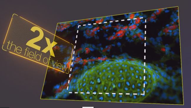
•diffraction limit relay lens supports 22mm FOV with large format sCOMS
•camera and spinning disk in conjugated image plane for maximum collection efficiency and easy alignment
•optical limit resolution confocal performance and beyond optical limit 120nm super-resolution performance
2. Excellent confocal effect from innovated spinning disk pattern and artefacts free:
•multi-spiral pinhole pattern ensures high fill factor and low pinhole cross-talk
•Non-isotropic pinhole arrangement allows higher spacing between pinholes and minimizes pinhole crosstalk from neighboring pinholes more efficient in covering the entire field of view rapidly in order of hundreds of microseconds to achieve 24 frames per rotation breakthrough from traditional 12 frames per rotation
•by-pass mode between wide field and confocal
•two pattern pinhole spinning disk and replacement “on the fly” with no alignment needed after changing the spinning
•disk box (spinning disk is well protected and sealed inside a box)to complement objective lens numerical aperture or any experiment protocol without limit
•pinhole disk pattern matching with EMCCD &sCMOS camera for high sensitivity or high speed image capture and super-resolution confocal structure illumination
•free of tailing artefacts in continuous 15,000RPM spinning under confocal mode
3. Continuous expansion flexibility to cope with existing requirements and future upgrade path:
•seamlessly integrate with existing microscope, even non-fluorescent manual microscope for multi-color 3D imaging
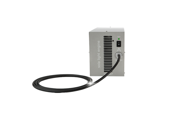
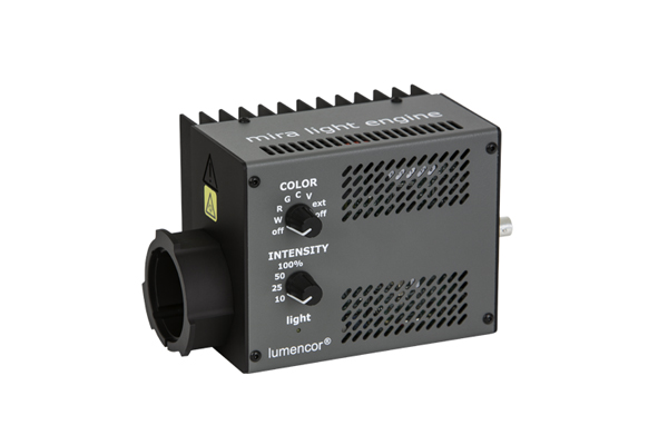
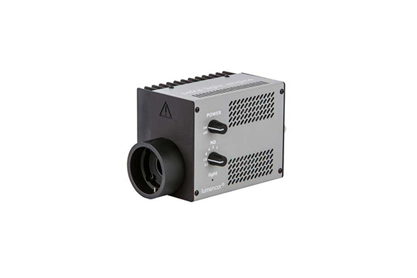
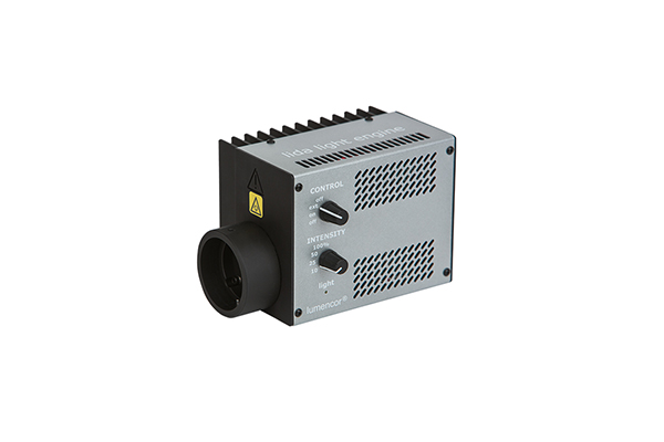
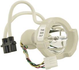
•choices of different high power long life light sources, high power LED (up to 310mW/λ) or high power laser (up to
•1W/λ in 1000:1 linear dynamic range)
•various software control package in continuous upgrade
•upgradeable super-resolution by structure illumination (both wide field and confocal mode)
•multi-cameras or camera image split options at any time
4. Reproducible high quality performance at all time from trouble free long term stability alignment
•alignment easy to adjust upon installation and stable for long time from the effective and neat optical design
•trouble free maintenance and operation
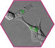
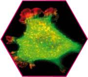
Left-side: Fluo-4 showing an induced Calcium signal, Right-side: Di-8-ANEPPS labeling the sarcolemma
Using 100X objective at 330fps capture speed, exposures at 2ms and streamed in a 200 frames
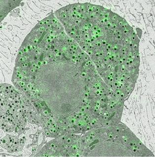
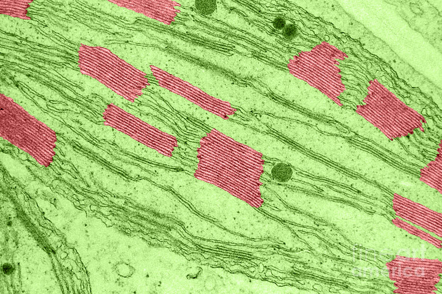
Chloroplasts moving inside fresh-water plant Elodea canadeus.
Z-Projection of 19 slices at 1um intervals; 10ms exposure of each slice and 500ms between 3D captures
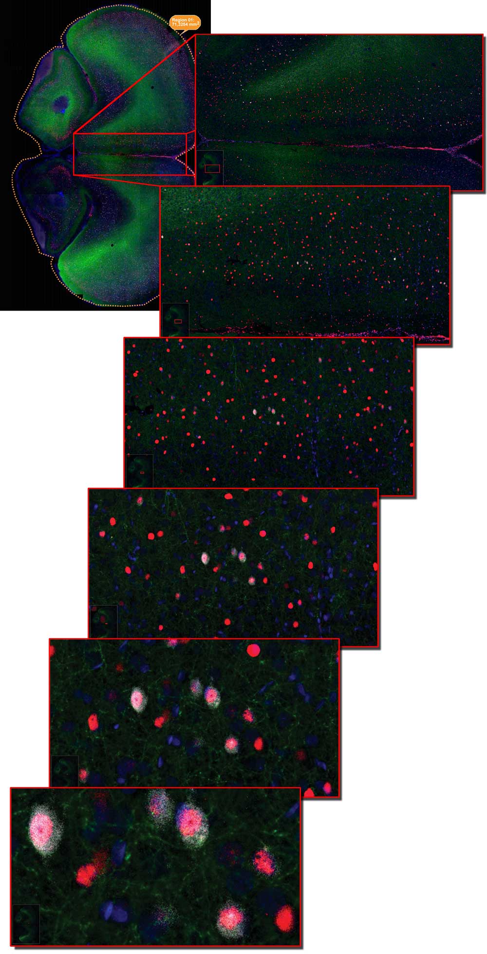
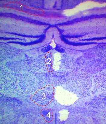
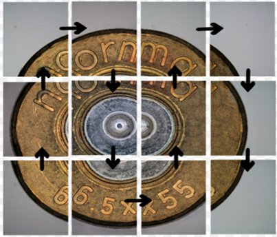
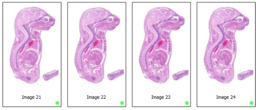
Mouse brain section confocal performance: scan a complete slice of mouse brain section with a size of 73,3mm2 with four channels and a 13 step Z-stack in 1,5 hours.
•60 μm thick section of an entire mouse brain sample is stained with 4 colors (3 antibodies and DAPI) and has been scanned by TissueFAXS Confocal at 20x magnification factor. 1400 images per each confocal plane and a total of 29400 images for 21 layers z-stack.
•Courtesy of TissueGnostics: acquisitions by courtesy of Janelia Farm Research Campus, Howard Hughes Medical Institute, Virginia, USA
•Note: TissueFAXS Confocal Plus SL integrates X-Light V2 scanner into the system
Top class spinning disk scanner ideal for living cell prolonged time lapse fluorescent imaging to monitor rapidly occurring events within living cells without compromising resolution, as well as the high frequency low intensity signals because of substantially reduced photobleaching and phototoxicity. Perfect for investigation of multicolor colocalization, 3D optical sectioning & reconstruction and Multi-dimension imaging (X, Y, Z, I, λ, t, n).
Specimen fluorescent staining: fluorescent dye staining, immuno-fluorescence assay, fluorescent in-situ hybridization, fluorescent protein, microinjection of fluorescent labeled actin probes into living cells.
Application areas:
•Cell Biology & Plant Biology: apoptosis, autophagy, cell cycle, cell metabolism, cell tracking and tracing, cytotoxicity, oxidative stress detection, phagocytosis, endocytosis, receptor internalization, cell signaling, & communication, cell motility, cellular compartmentalization, protein synthesis and degradation, cellular and biophysical regulations,
•Cell and System Dynamics: structures and organs, e.g. blood vessels, neurons and processes such as angiogenesis and immune responses to vessel lesions
•Embryology & Developmental biology: C. elegans, Drosophila and Zebrafish growth and signal mechanisms
•Cancer research
•Clinical & translational medicine research
•Cardio and neuro sciences
•Calcium imaging, other ion measurements & membrane potential
•Yeast and bacteria studies
•Stem cell research and 3D cultures
•Synthetic biology: biofuels, vaccine & antibody production, plant sciences, industrial enzymes, biobased chemicals
•Pharma, biopharma & CRO
Crest Optics X-Light V2 Specification | |
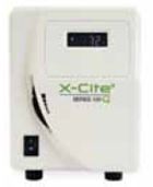
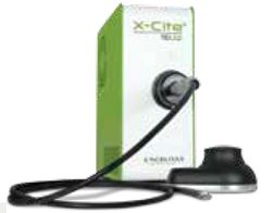
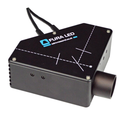
Light source | Flat and even illumination by LED (3 to 7 wavelengths) or Laser (3 to 6 modules or 6 or 7wavelengths combination) with superb rejection of out of focus light |
Illumination source supports hardware triggering for fast multichannel experiment | |
Supported input fiber | 0.39NA multimode 1.5mm fiber with SMA adapter; excitation Gimbal mount for easy alignment on custom microscope setup and for best S/N |
Acquisition modes | Widefield – disk out, capture bright field phase contrast or DIC overlay with confocal image or standalone in all wide field fluorescent & bright field imaging |
Confocal – disk in | |
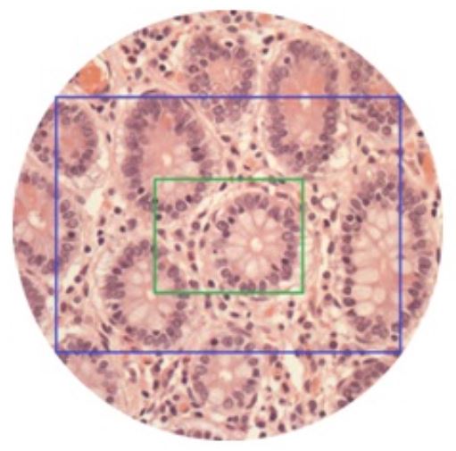
Disk pinhole size vs camera size | Double pattern pinholes at 12mmx12mm FOV each pattern for CCD |
Single pattern pinholes at 16.8mmx14mm FOV for large format sCMOS | |
Option: custom pattern | |
Disk speed | 15,000 RPM =6,000fps |
Disk changing | Hot-swap the spinning disk box with no alignment needed after changing the spinning disk box for maximum pinhole pattern flexibility in the same confocal system (spinning disc is well protected and sealed inside a box) |
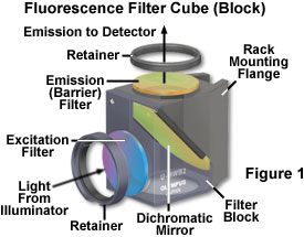
Laser clean up filter | Manual 4-position slider (option 8-position motorized filter wheel) |
Dichroic wheel | Motorized 5-position standard |
Dichroic size | 25.5 mm x 36 mm x 1 mm |
Emission filter wheel | Automated 8-position wheel standard |
Emission filter size | Φ 25 mm |
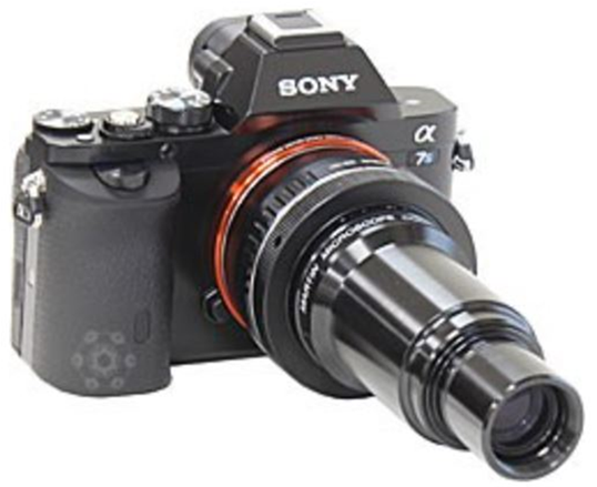
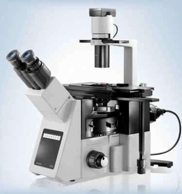
Supported microscope | Upright and inverted microscope models from all brands with 100% c-mount output port |
With apochromatic corrected objective | |
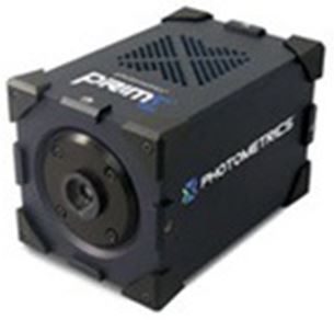
Camera | High sensitivity CCD, sCMOS and EMCCD in c-mount port |
Easy camera focus through internal optics without moving camera, no other disc and camera alignment needed | |
Software control | MetaMorph, NIS Elements, Micro-Manager |
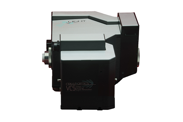
VCS | Compatible and upgradeable with X-Light V2 scanner |











 关闭返回
关闭返回